Sunday, June 22, 2014

Making Blood Culture Workups More Efficient

A major problem with blood culture workups is that it is often difficult to determine a true positive (pathogen) result from a false positive (contaminant) result. One solution is to adjust the testing algorithm in place. In 1970, it was shown that 11% of contaminated cultures had additional positive cultures, while 69% of clinically significant cultures had multiple positive cultures. In the proposed algorithm (summarized right), any
isolates that are typically contaminants that are not present in follow-up cultures are reported as contaminants. To confirm the accuracy of the new algorithm, a nurse epidemiologist and an infectious diseases physician reviewed the patient charts.

Overall, organisms considered to be contaminants (CNS, diphthroids, Micrococcus spp, Bacillus spp, and viridans streptococci) were identified in 495/1040 positive blood cultures. Of the 495, 286 were classified as contaminants due to negative follow-up cultures, 171 were investigated by a pathology resident, and 15 were classified as pathogens (viridans strep with positive follow-up cultures). The majority (62%) of the contaminants were identified as CNS.

Two types of errors, VM (very major) and M (major) were used to determine the success of the new algorithm. A "very major error" occurred if an isolate that was classified as a contaminant was determined to be clinically significant after chart review. A "major" error occurred if an isolate was classified as indeterminate or pathogenic and was later determined to be not clinically significant. VM errors occurred in 6.3% of cases and M errors occurred in 6.6% of cases. Further adjustment of this algorithm may decrease the error rate in the future, but the algorithm was determined "acceptable" by the authors of this study.

Source: http://jcm.asm.org/content/40/7/2437.full

Friday, June 13, 2014

PED Virus Increasing Pork Prices

In May 2013, porcine epidemic diarrhea virus (PEDv) was isolated in hog herds in the United States. PEDv cause diarrhea and vomiting in pigs (morbidity near 100%) with variable mortality. Older pigs are more likely to survive the virus than young pigs (mortality 50-100%). The virus has an incubation period of 3-4 days, and is spread more slowly than other types of swine gastroenteritis. Pigs who recover do so within 7-10 days of symptom onset. The virus is spread through a fecal-oral route, but fomites and contaminated equipment may spread the virus as well.

PEDv was first reported in the United Kingdom in 1971, and has spread to several countries since then.
PEDv is a Coronavirus, containing an enveloped ssRNA genome. The virus does not appear to have jumped to humans in the 40 years since its discovery.

Global AgriTrends speculates that up to 4.5% of pigs, or 4.5 million hogs, in the US may be killed by PEDv this year. Some farms have lost 10% of their herds already. Pork prices are expected to increase 3% this year, compared to 0.9% last year. A vaccine is not yet available for the virus in the US, but there are vaccines currently available in South Korea, Japan, and China.

Sunday, June 8, 2014

Bacteria Cultured from a Bear Bite

One of the dangers of an animal bite (aside from the trauma of the bite itself) is the possibility of infection due to flora in the mouth of the animal. If an infection does develop from an animal bite, knowing the flora present is helpful in choosing antibiotics that will be effective. Most animals that interact with humans (cats, dogs, etc) have well-defined flora. Less is known about other animals, such as bears, that rarely bite humans.

A case report from 2004 describes the bacteria cultured from a bite wound inflicted on a hunter by a grizzly bear in Canada. Cultures taken about 12 hours after the bite grew Serratia fonticola, Serratia
marcescens (used to draw a raptor, right), Aeromonas hydrophila, Bacillus cereus, and Enterococcus durans. No anaerobes were isolated from the wound. The patient was given 1 week of piperacillin-tazobactam therapy followed by  amoxicillin-clavulanate and ciprofloxacin for 3 weeks as prophylactic treatment.

Infection after a bear bite is common, with around 44% of patients showing infection. However, few of these bite wounds have been cultured for identification of flora, so there is limited knowledge of what is common or what antimicrobials are appropriate for treatment. Some scientists hypothesize that oral flora of bears will be similar to that of dogs, as well as being highly variable depending on the foraging environment and habits of a particular bear.

More information about the patient and his clinical outcomes can be found here: http://www.ncbi.nlm.nih.gov/pmc/articles/PMC446265/#__ffn_sectitle

Saturday, May 31, 2014

Malaria Vaccine in the Works

Malaria is a disease caused by parasitic organisms of the Plasmodium family that are transmitted by the bites of infected Anopheles mosquitoes. Of the four species that cause illness in humans, P. falciparum is the most common and the most deadly. Malaria is most common in Africa, but is present throughout the world. Malaria is an acute febrile illness, with symptoms such as fever, nausea, or vomiting appearing 10-15 days after infection. If treatment is not begun within 24 hours, P. falciparum malaria can cause severe illness or even death.

A new vaccine for malaria is currently in early stage trials with promising results! The vaccine was developed after a survey of Tanzanian children found that 6% of them had antibodies to a protein crucial to the parasite's development. The antibody prevents the parasite, Plasmodium falciparum (picture right: organisms in intracellular phase), from reproducing within the blood cell. This allows the spleen and macrophages time to remove the damaged cells from circulation. The children with the antibody did not suffer from severe malaria, consistent with a previous study in Kenyan adolescents. This new vaccine is one of around 100 vaccines that have been developed since 1980, but is the first to focus on the parasite once it has entered the body.


Picture: http://lib.jiangnan.edu.cn/ASM/257-1.jpg
References
http://www.forbes.com/sites/paulrodgers/2014/05/23/has-malaria-met-its-match/
http://www.who.int/mediacentre/factsheets/fs094/en/

Friday, May 23, 2014

Lemierre's Syndrome: A Case Report

Lemierre's Syndrome (also known as postanginal septicemia and human necrobacillosis) was first described by French physician and microbiology professor Andre Lemierre in 1936. He classified this syndrome as an "anaerobic postangial sepsis" caused primarily by  Bacillus funduliformis (known today as Fusobacterium necrophorum), a gram-negative anaerobe (organism on SBA, right). This organism is responsible for 90% of Lemierre's syndrome cases and 21% of recurrent sore throats.

Before the introduction of antibiotics, Lemierre's syndrome had a mortality rate around 90%. Currently, the mortality rate of correctly-diagnosed cases is around 5%. Fewer than 100 cases have been described since 1974, and the condition has a incidence rate of 3.6 cases per 1 million. This syndrome is characterized by a recent history of oropharyngitis with persistent fevers, followed by septic thrombophlebitis (swelling of a vein caused by a blood clot) of the internal jugular vein as well as dissemination of the infection to multiple sites. Lemierre's syndrome should be suspected in patients who present with pharyngitis and high-grade fevers (39-41C) plus thrombophlebitis or sepsis.


Case Report
A previously healthy 44 year old female presented with a 3 day history of worsening sore throat predominantly on the left side with fever, pain while swallowing, and vomiting. Upon examination, she was febrile, hypotensive, and tachycardic. Her left tonsil was displaced, and her lymph nodes were swollen and tender. Laboratory testing showed her WBC to be elevated with a decreased platelet count, as well as decreased liver and kidney function. A CT scan (right) showed a mass in the left peritonsillar region (arrow). Drainage of the mass was delayed due to the patient's unstable condition. The patient deteriorated rapidly, developing acute renal failure, encephalopathy, respiratory failure, and bilateral pleural effusions. She was placed on mechanical ventilation and dialysis. Blood cultures grew F. necrophorum and the patient's antibiotics were adjusted to piperacillin-tazobactam and metronidazole. The patient was extubated on her ninth day of admission, and discharged on the thirteenth.

References
SBA picture: http://pictures.life.ku.dk/atlas/microatlas/veterinary/bacteria/Fusobacterium_necrophorum_A/fusobacteriumnecrophoruma.jpg
Case Study:  : Arora T, Wright D (2014) Lemierre’s Syndrome in an Adult: A Case Review. Otolaryngology 4: 167. doi:10.4172/2161-119X.1000167

Friday, May 16, 2014

Ebola!


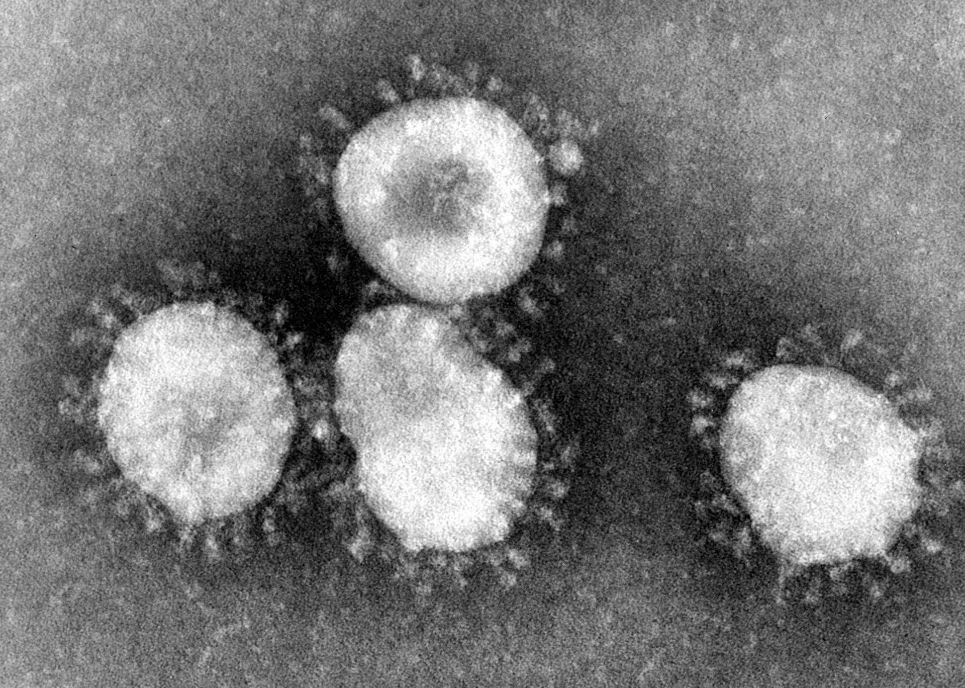
Ebola (pictured right) is an RNA filovirus in the family Filoviridae, genus Ebolavirus. Ebola was first discovered in 1976 in the Democratic Republic of the Congo. Of the five species, four cause serious disease in humans and one only causes disease in nonhuman primates. Ebolavirus is spread through direct person-to-person (or primate-to-primate) contact with blood, secretions, organs or bodily fluids of infected patients.

Ebolavirus infection is fatal in 50-90% of cases, with some strains less virulent (50-60%) and others more virulent (80-90%). Symptoms appear 2-21 days post-exposure with a rapid onset. Initial symptoms are flu-like, but quickly progress to more serious symptoms such as chest pain, red eyes, skin rash, jaundice, hiccups, or bleeding. Laboratory findings of interest are low WBC/platelet counts with elevated liver enzymes. Definitive diagnosis of ebolavirus occurs through ELISA, antigen detection or serum neutralization tests, RT-PCR assays, electron microscopy, or viral culture.

The virus produces proteins that increase blood vessel permeability, causing hemorrhage. Ebolavirus may also prevent the body from mounting an appropriate immune response through an unknown mechanism. One theory is that the virus overwhelms the immune system using a cytokine storm, sending the patient into shock. Another theory is the virus prevents the immune system from mounting a response at all by reducing interferon activity within the cells (normally, this activity would signal NK cells or T cells that a cell has been taken over by a virus and mark it for destruction).

Unfortunately, there is no treatment for ebolavirus, but a vaccine is in clinical trials. Infected patients are placed into quarantine and receive supportive therapy, such as pain medicine or fluids. Death results from pulmonary or gastrointestinal hemorrhage, hepatitis, or encephalitis one to two weeks after the onset of symptoms. Patients who recover may remain infectious for several weeks after symptoms clear.

References
Picture: http://edwardmd.files.wordpress.com/2013/11/ebola1.jpg
1. Basler Christopher, et al. The Ebola Virus VP35 Protein Inhibits Activation of Interferon Regulatory Factor 3. Journal of Virology. July 2003: 88(11).
2. CDC. Ebola Hemorrhagic Fever. CDC Viral Hemorrhagic Fevers. 2014. Available at: http://www.cdc.gov/vhf/ebola/. Accessed May 16, 2014.
3. Federation of American Scientists. Ebola Fact Sheet. FAS Biosecurity Fact Sheets. Available at: http://www.fas.org/programs/ssp/bio/factsheets/ebolafactsheet.html. Accessed May 16, 2014.
4. Villinger Francois, et al. Markedly Elevated Levels of Interferon (IFN)-y, IFN-a, Interleukin (IL)-2, and Tumor Necrosis Factor-a Associated with Fatal Ebola Virus Infection. The Journal of Infectious Diseases. 1999: 179 pp S188-S191.
5. WHO. Ebola virus disease. WHO Media Centre Fact Sheets. 2014. Available at: http://www.who.int/mediacentre/factsheets/fs103/en/. Accessed May 16, 2014.

Tuesday, May 6, 2014

Welcome!

Hello blog readers and welcome to my blog, Microbi-blog-ogy.

I am a graduate student in UAB's CLS program, and my interests include bluegrass music, duathlon/triathlon, and crossword puzzles. My favorite color is purple, and my favorite movie is O Brother, Where Art Thou?. I received my Bachelor's degree in Laboratory Technology from Auburn University in 2013. My skills include LaTeX, time management, computer applications, and accepting visual, auditory, and tactile inputs and translating them into kinesthetic outputs

Currently, I am taking an infectious diseases course as part of my Master's degree program. This blog will showcase topics of interest that I encounter throughout this course.